Please hover over the image below to explore more detail. Additional information can be discovered by scrolling down.


SID Interactive Skin Model
The Society for Investigative Dermatology (SID) is pleased to unveil a new interactive skin model that highlights the biology on which our Society is founded.
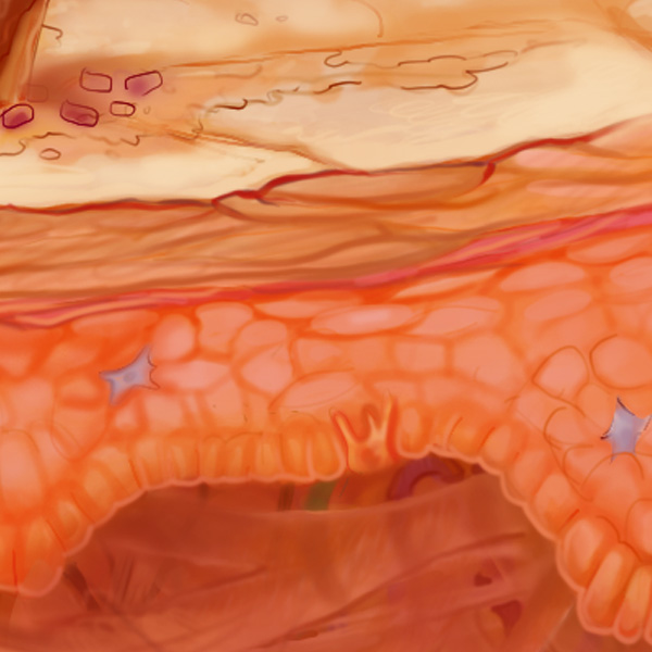
With few available images that capture the key elements of skin structure, the SID has partnered with Digizyme, a Massachusetts-based scientific visualization company, and collaboratively created what we feel is a wonderful depiction that progresses from macro on the left to more finely detailed on the right.
With an eye toward future applications, we intend to use this image for educational purposes and as a conduit to SID-associated research addressing structures within the image. To capture more aspects of the skin at macro, micro, and ultrastructural levels, we crafted layers of the drawing to allow flexibility regarding potential future applications. Options will include animations of relevant biology of the various aspects of skin and its development.
This image will be featured in SID’s collateral offerings and website going forward. We are excited to share this project with our SID membership and anticipate it will be useful for skin biology and educational communities more broadly.
This image will evolve through feedback over time. If you have comments or suggestions please send them to us at sid@sidnet.org.
Please check back to track the evolution of the SID’s skinteractive model!
SID Skin Model Printable Materials
SID is pleased to provide a print quality version of our interactive skin model, allowing for the model to be used as a learning tool in printable formats for educational settings around the world.
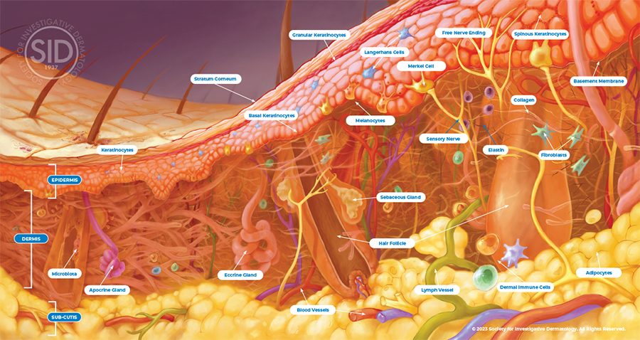
Skin Model Definitions
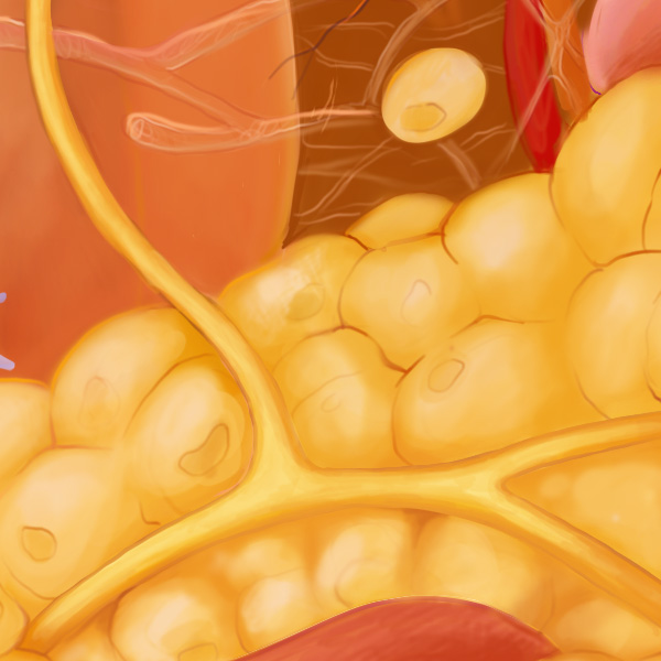
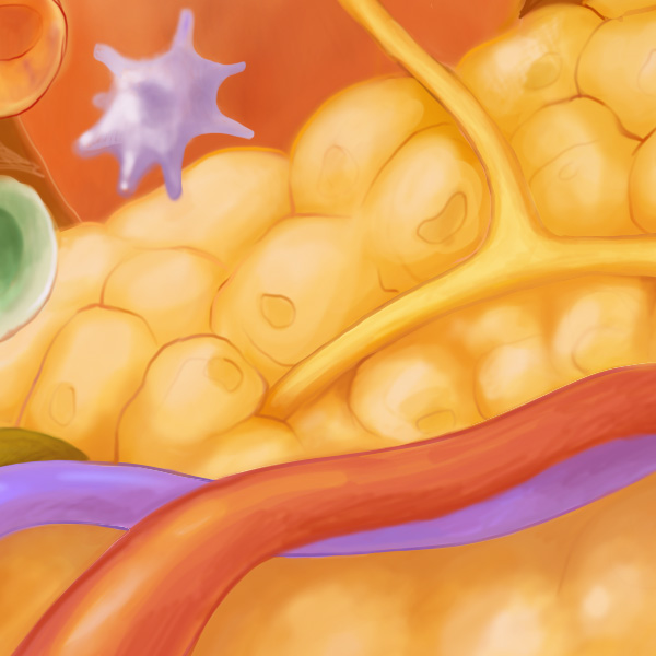
An adipocyte is a type of fat cell that can accumulate into many small pockets of fat to form adipose tissue. Adipose tissue helps to insulate the body and store energy and can also release hormones and other substances that can affect the body’s metabolism and other functions. Adipocytes are located in the sub-cutis, just below the dermis.
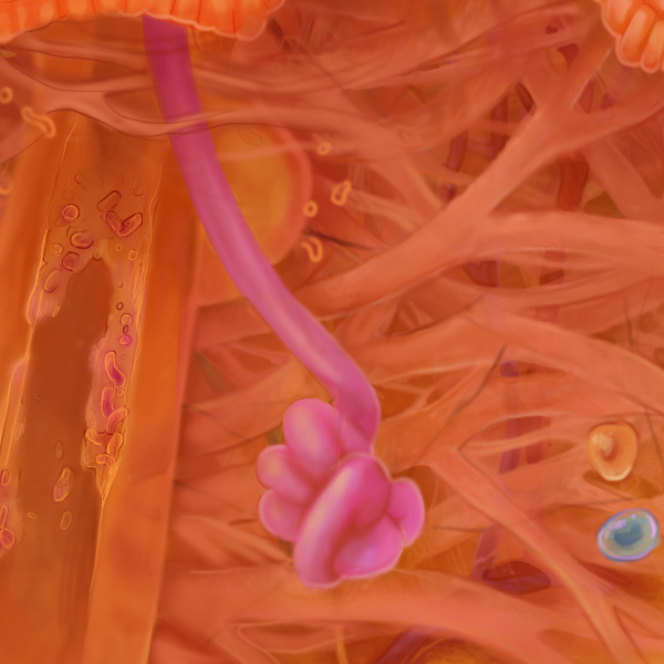
Apocrine glands are composed of a group of cells specialized for the release of secretions. They are mostly located in the axillae and groin. The secretions are odorless until acted on by micro-organisms.
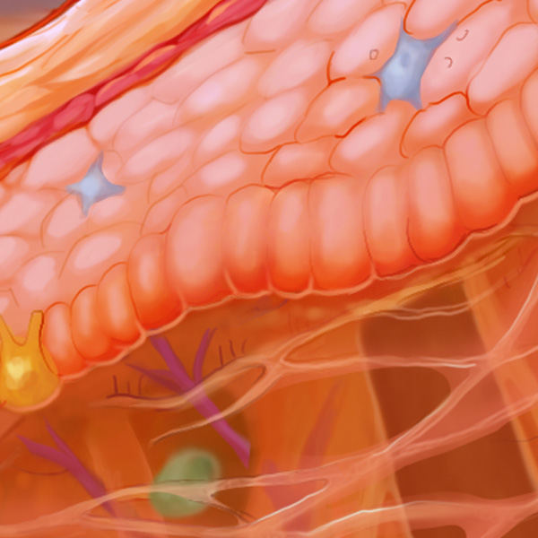
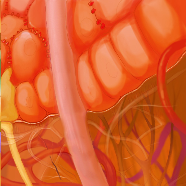
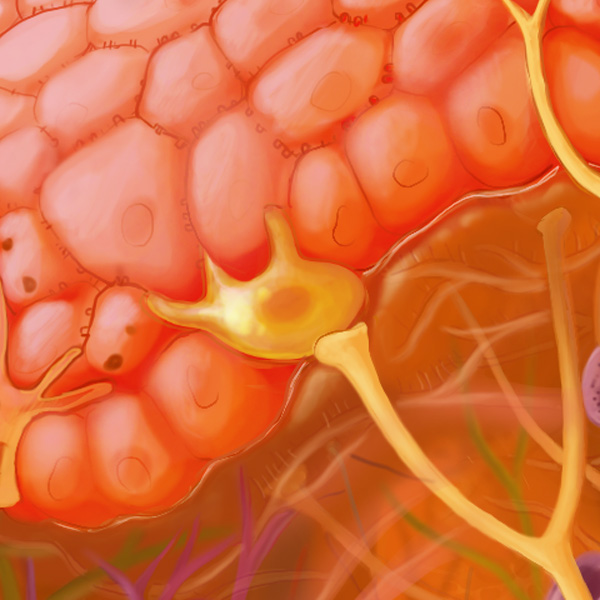
Basal keratinocytes are at the bottom (basal) layer of the epidermis. These cells constantly divide, migrate towards the surface of the skin, and produce keratin — a protein that protects the skin and affects its appearance. Keratin makes human skin smooth and soft, but when produced in excess can result in rough calluses or general scaliness.
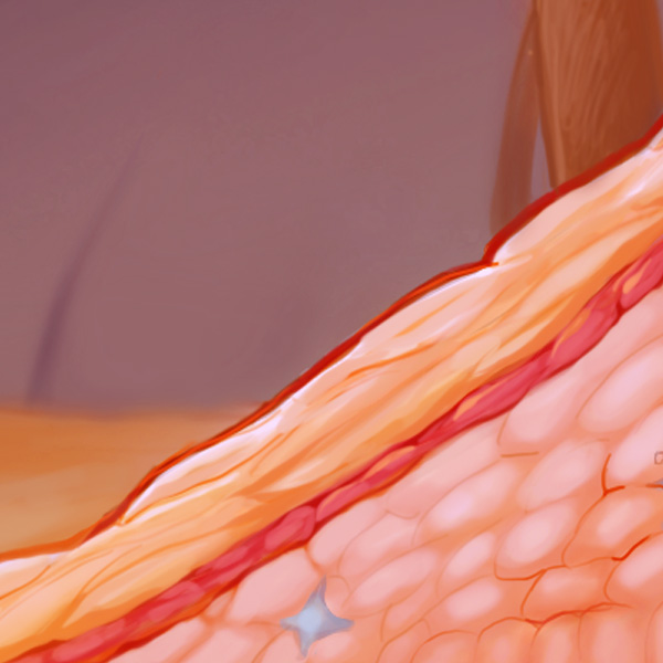
The basement membrane is a thin, strong layer of tissue that acts like a foundation for cells in the body. It helps to keep different types of tissue separate, supports the cells that are on top of it, and helps play a role in the movement of substances between cells. The basement membrane also acts as a barrier that inhibits tumor invasion of deeper tissue layers.
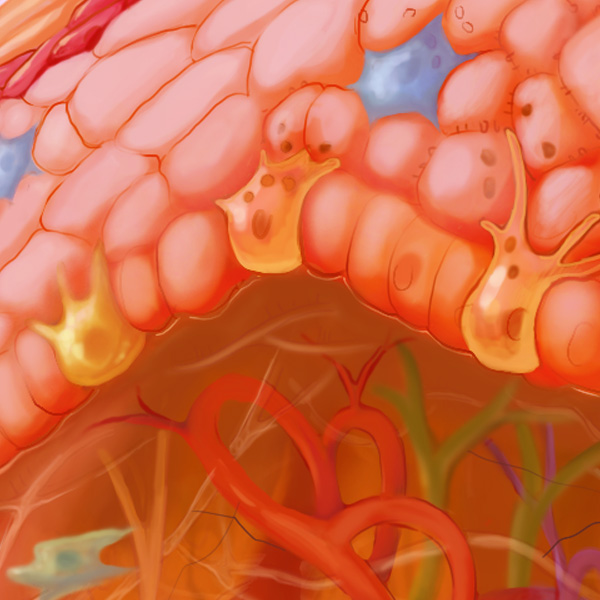
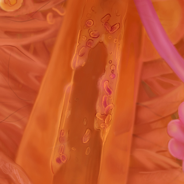
Blood vessels circulate blood cells, nutrients, and oxygen to the tissues of the body. They also take waste and carbon dioxide away from the tissues. In the skin, small blood vessels penetrate to the top of the dermis, but the epidermis is not vascularized and receives it nutrients from across the dermal-epidermal junction.
Collagen is a protein that gives structural integrity and strength to various tissues in the body, including skin, tendons, ligaments, and bone. It is also involved in wound healing and tissue repair. Collagen is the most abundant protein in the human body, making up over 30% of the body’s total protein!
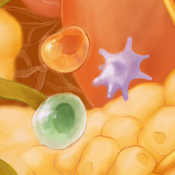
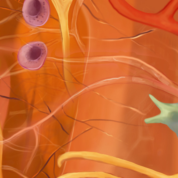
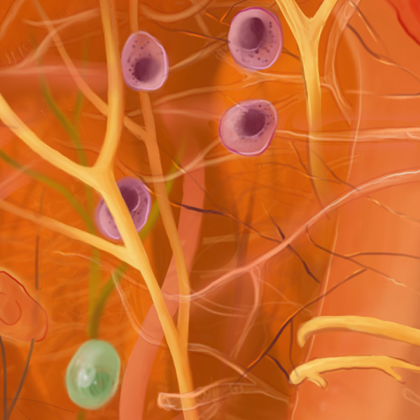
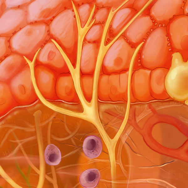
Dermal immune cells are found in the dermis, the middle layer of the skin, and help protect the body from infection and disease by recognizing and attacking harmful substances that enter the body through the skin. Dermal immune cells include a variety of cell types, including T cells, B cells, and dendritic cells.
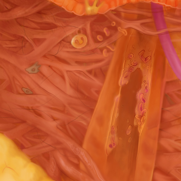
The dermis layer of skin lies beneath the epidermis, for which it provides support and nourishment. It is made up of strong flexible connective tissue and contains blood vessels, nerve endings, sweat glands, and hair follicles. The dermis plays a role in regulating body temperature, sensation, and production of substances including sweat and oil.
Eccrine sweat glands are the major source of sweat for the human body. They are found in virtually all skin, with the highest density in palms and soles, then on the head, but much less on the torso and the extremities. Eccrine glands are found in other mammals, but mainly in the palms and soles of feet to provide grip.
Elastin is a protein that helps tissues maintain their elasticity and flexibility. It is found in tissues such as skin, blood vessels, and lung, and helps them return to their original shape after being stretched or bent, preventing tearing or damaging of the tissue. Wrinkles develop in part due to a loss of elastin in the skin.
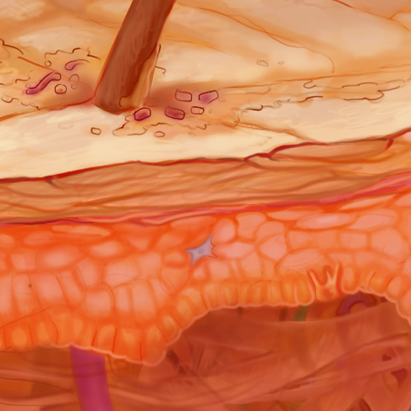
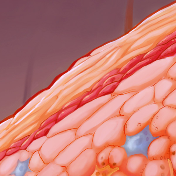
Constantly shedding and renewing itself, the epidermis is made up of several layers of cells that form the outermost layer of skin. The epidermis is responsible for protecting the body from external harm such as bacteria, ultraviolet radiation, and physical trauma. It also helps to maintain the body’s temperature and hydration levels.
Fibroblasts are cells found in the connective tissues throughout the body. They produce and maintain collagen and elastin, structural proteins which give strength and elasticity to tissues. Fibroblasts also play a role in wound healing by producing new collagen and other extracellular matrix components which help repair and rebuild damaged tissue.
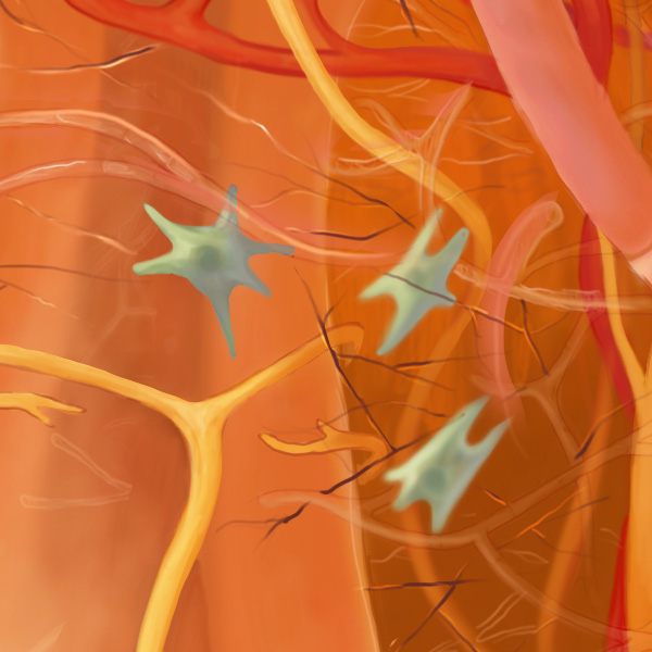
Free nerve endings are simple, branching fibers that can detect various types of stimuli (such as pain, temperature, touch) and send these signals to sensory nerves. They are unencapsulated, meaning they do not have a specialized structure surrounding them like other types of sensory nerve endings and are not dedicated to any specific sensation.
Granular keratinocytes are a type of keratinocyte named for the granules of keratin that they contain, which are visible under a microscope. Granular keratinocytes are terminally differentiated keratinocytes, which means they can no longer change into a different type of keratinocyte. These are found in the granular layer of the skin.
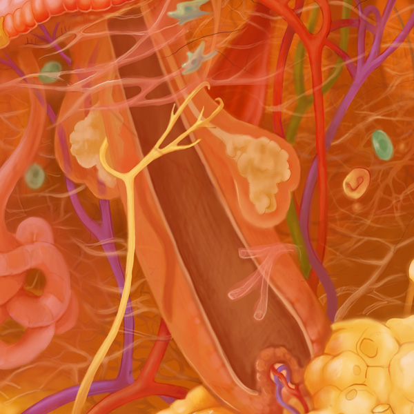
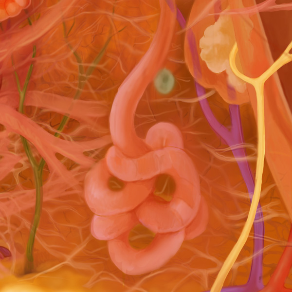
Hair follicles reside in the dermal layer of the skin and regulate hair growth via a complex interaction between hormones, neuropeptides, and immune cells. These interactions induce hair follicles to produce different types of hair as seen on different parts of the body. It is estimated that the average human scalp has 100,000 to 150,000 hair follicles.
Keratinocytes are a type of cell that is found in epidermis. They are responsible for producing a protein called keratin, which protects and keeps the skin strong. As keratinocytes mature, they migrate towards the skin’s surface, where they eventually die and form a protective barrier.
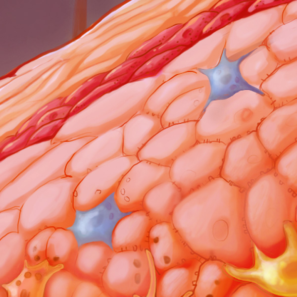
Langerhans cells (LC) form a dense network of immune system “sentinels” in the epidermis. LCs determine which substances should be tolerated versus attacked by T cells. When LCs detect a “dangerous” substance, they act as antigen-presenting cells. They can be found in other tissues, such as lymph nodes, and are associated with Langerhans cell histiocytosis (LCH), which is a rare disorder that causes tissue damage when there are too many abnormal LCs.
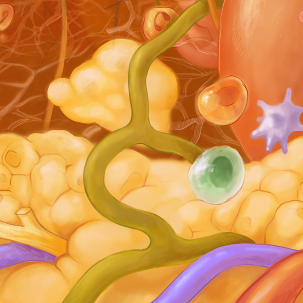
Lymph vessels are thin-walled tubes that carry lymph, a fluid made up of white blood cells and other infection-fighting substances, back from tissues toward the heart. Groups of lymphocytes organize around lymph vessels to form lymph nodes (which swell with accumulated lymphocytes when you’re sick!).
Melanocytes are located in the bottom layer of the epidermis and produce melanin, a pigment which gives a brownish color to the skin, hair, and eyes. Melanocytes protect the skin against ultraviolet radiation by increasing melanin production to shield the DNA (a process known as tanning). Melanocytes are the cell of origin for the aggressive skin cancer, melanoma.
Merkel cells are a type of nerve cell found in the epidermis, responsible for sensing light touch and pressure. They are often found in clusters near the base of hair follicles and are highly abundant in especially sensitive skin areas such as the fingertips and lips. A rare, aggressive skin cancer called Merkel cell carcinoma (MCC) arises from uncontrolled growth of cells that share certain characteristics with normal Merkel cells.
Skin flora, also called skin microbiota, refers to micro-organisms that reside on the skin. Most are found in the superficial layers of the epidermis and the upper parts of hair follicles. Their many interactions with the skin are actively being investigated, but can be beneficial, neutral, or detrimental to the host. There are around 1,000 species of bacteria that can inhabit human skin, belonging to nineteen phyla.
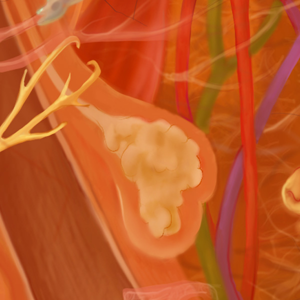
Sebaceous glands produce sebum, an oily, innately-antibacterial substance which keeps the skin moist. These glands are located all over the body, with the highest density in the face and scalp. Clogged sebaceous glands can contribute to several skin conditions including acne, rosacea, and seborrheic dermatitis.
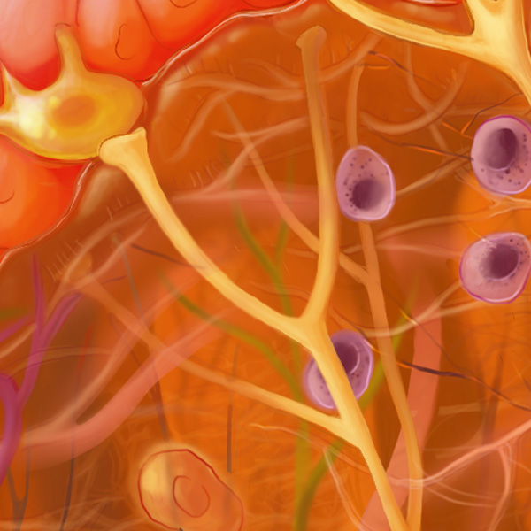
Sensory nerves are responsible for transmitting sensory information from the body to the central nervous system to help perceive the environment. This includes information about touch, temperature, pain, and other sensations. Sensory nerves carry signals from sensory receptors throughout the body and travel to the spinal cord and the brain where the information is processed.
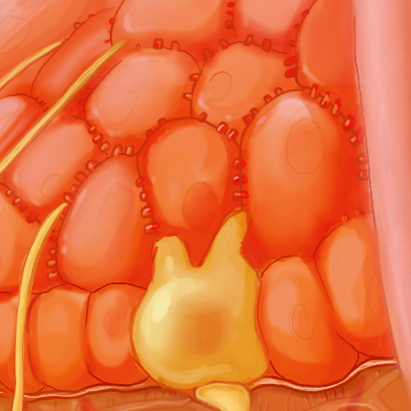
Spinous keratinocytes are a subtype of keratinocyte named for their spiky, spiny appearance under a microscope, due to the presence of long, thin processes called tonofilaments which anchor the cytoskeleton to the cell membrane. Spinous keratinocytes are located in the spinous layer of the epidermis, between the basal and granular layers.
The stratum corneum is the outermost layer of the epidermis and the final differentiated form for keratinocytes in the skin. It consists of several layers of flattened cells called corneocytes. Constantly sloughing off (forming the major food source for dust mites in your home!), the stratum corneum is a “dead” tissue that forms a barrier to protect the body from infection, dehydration, chemicals, and mechanical stress.
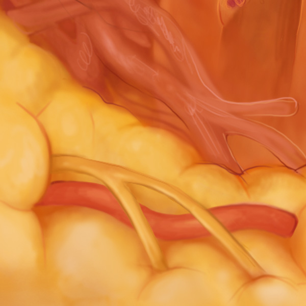
Below the dermis, the sub-cutis consists mostly of fatty cells and provides cushioning and energy storage.